-
Collagen
-
Type I - Atelocollagen
- PureCol® Solution, 3 mg/ml (bovine) #5005
- Nutragen® Solution, 6 mg/ml (bovine) #5010
- FibriCol® Solution, 10 mg/ml (bovine) #5133
- PureCol® EZ Gel, Solution, 5 mg/ml (bovine) #5074
- PureCol® Lyophilized, 15 mg (bovine) #5006
- VitroCol® Solution, 3 mg/ml (human) #5007
- VitroCol® Lyophilized, 15 mg (human) #5008
-
Type I - Telocollagen
- TeloCol®-3 Solution, 3 mg/ml (bovine) #5026
- TeloCol®-6 Solution, 6 mg/ml (bovine) #5225
- TeloCol®-10 Solution, 10 mg/ml (bovine) #5226
- RatCol® for 3D gels, Solution, 4 mg/ml (rat) #5153
- RatCol® High Concentration, Solution, 10 mg/ml (rat)
- RatCol® lyophilized, 100 mg (rat)
- RatCol® for Coatings, Solution, 4 mg/ml (rat) #5056
- Type I - Insoluble Collagen
- Type I - Bioinks
- Type II Collagen
- Type III Collagen
- Type IV Collagen
- Collagen Standard
- PureCol® Collagen Coated Plates
- Collagen Scaffolds
- Collagen Hybridizing Peptides
-
Type I - Atelocollagen
- Tunable Stiffness
- CytoSoft® Rigidity Plates
-
Bioprinting
- Support Slurry for FRESH Bioprinting
- Collagen Bioinks for Extrusion Bioprinting
- GelMA Bioinks for Extrusion Bioprinting
- Photoinitiators
- Bioinks and Components for DLP Bioprinting
- Bioink Components
- Methacrylated Collagen
- Methacrylated Gelatin
- Methacrylated Hyaluronic Acid
- Diacrylates
- Methacrylated Polysaccharides
-
3D Hydrogels
- Thermoreversible Hydrogel
- Silk Fibroin
-
Type I Collagen for 3D Hydrogels
- PureCol® Solution, 3 mg/ml (bovine) #5005
- Nutragen® Solution, 6 mg/ml (bovine) #5010
- FibriCol® Solution, 10 mg/ml (bovine) #5133
- PureCol® EZ Gel, Solution, 5 mg/ml (bovine) #5074
- VitroCol® Solution, 3 mg/ml (human) #5007
- TeloCol®-3 Solution, 3 mg/ml (bovine) #5026
- TeloCol®-6 Solution, 6 mg/ml (bovine) #5225
- TeloCol®-10 Solution, 10 mg/ml (bovine) #5226
- RatCol® for 3D gels, Solution, 4 mg/ml (rat) #5153
- HyStem® Thiolated Hyaluronic Acid
- Methacrylated Collagen
- Methacrylated Gelatin
- Methacrylated Hyaluronic Acid
- Diacrylates
- Collagen Sponges
- Methacrylated Polysaccharides
- Extracellular Matrices
- HyStem / Hyaluronic Acid
-
Adhesion Peptides / Proteins
-
Recombinant Adhesion Proteins
- CD2, 0.5 mg/ml #5086
- CDH3, 0.5 mg/ml #5124
- CDH13, 0.5 mg/ml #5125
- CD14, 0.5 mg/ml #5089
- CDH18, 0.5 mg/ml #5090
- CD40, 0.5 mg/ml #5093
- CD86, 0.5 mg/ml #5096
- CD164, 0.5 mg/ml #5100
- CD270, 0.5 mg/ml #5127
- CD274, 0.5 mg/ml #5126
- CD276, 0.5 mg/ml #5123
- E-Cadherin (CD324), 0.5 mg/ml #5085
- ICAM2, 0.5 mg/ml #5107
- Adhesion Peptides
- Collagen Hybridizing Peptides
-
Recombinant Adhesion Proteins
- Reagents
- Assays
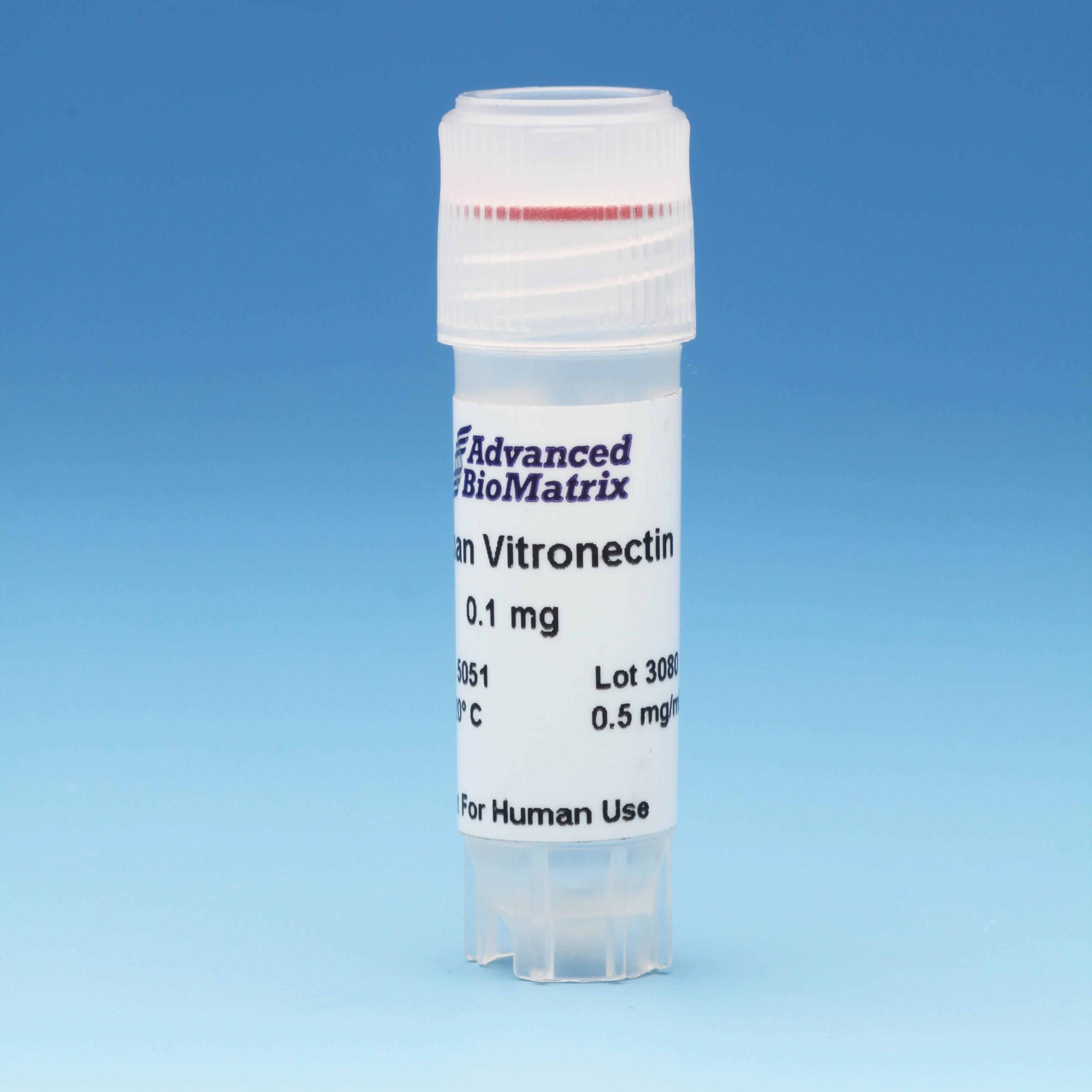
Vitronectin
Solution, 0.5 mg/ml (Human)
Catalog #5051
Vitronectin
Solution, 0.5 mg/ml (Human)
Catalog #5051
This Vitronectin solution has been purified from human plasma where it is found as a mixture of 75kDa and 65kDa polypeptides. Vitronectin’s primary use in cell culture is related to cell adhesion. It also binds to heparin and collagen.
Product Description
Vitronectin is a monomeric glycoprotein used to promote cell attachment, migration, proliferation and differentiation in a broad number of cell lines and types. This product has been purified from human plasma where it is found as a mixture of 75kDa and 65kDa polypeptides.
Vitronectin’s primary use in cell culture is related to cell adhesion. It also binds to heparin and collagen.
Vitronectin is ideal for coating of surfaces. The optimal concentration for cell attachment and culture may differ for various cell types. Vitronectin has been used at a final coating concentration as low as 50 ng/cm2 on plasticware. It is provided in user-friendly packaging for use and storage. Vitronectin is sterile filtered and is supplied as a ready to use solution after thawing and concentration adjustment. This product is shipped separately on dry ice.
| Parameter, Testing, and Method | Vitronectin #5051 |
| Quantity | 0.1 mg |
| Volume | 0.2 mL |
| Concentration | 0.5 mg/mL |
| Purity - SDS PAGE Electrophoresis | >95% |
| Formulation | 0.15 M NaCl, 0.005M HEPES pH 7.4 |
| Form | Solution |
| Source | Human, Plasma |
| Storage Temperature | -20°C or -70°C for long term storage |
| Shelf Life | Minimum of 6 months from date of receipt |
| Sterilization Method | Filtration |
| Cell Attachment Assay | Passes |
| Sterility - USP modified | No growth |
| Safety | Source material found negative for infectious agents |
Directions for Use
Download the full PDF version or continue reading below:
Coating Procedure:
Use these recommendations as guidelines to determine the optimal coating conditions for your culture system.
- Thaw Vitronectin and dilute to desired concentration using serum-free medium or PBS . The final solution should be sufficiently dilute so that the volume added covers the surface evenly.
- Add appropriate amount of diluted material to culture surface.
- Incubate at room temperature for approximately 1 – 2 hours.
- Aspirate remaining material.
- Rinse plates carefully with dH2O– avoid scratching bottom surface of plates.
- Plates are ready for use. They may also be stored at 2-8°C damp or air dried if sterility is maintained.
Product References
Vitronectin References:
Wiley, Luke A., et al. "Using patient-specific induced pluripotent stem cells and wild-type mice to develop a gene augmentation-based strategy to treat CLN3-associated retinal degeneration." Human gene therapy 27.10 (2016): 835-846.
Wiley, Luke A., et al. "Generation of xeno‐free, cGMP‐compliant patient‐specific iPSCs from skin biopsy." Current protocols in stem cell biology 42.1 (2017): 4A-12.
Dwyer, Sheila Figel, Lingqiu Gao, and Irwin H. Gelman. "Identification of novel focal adhesion kinase substrates: Role for FAK in NFκB signaling." International journal of biological sciences 11.4 (2015): 404.
Wiley, Luke A., et al. "cGMP production of patient-specific iPSCs and photoreceptor precursor cells to treat retinal degenerative blindness." Scientific reports 6 (2016): 30742.
Kittur, Harsha, et al. "Probing Cell Adhesion Profiles with a Microscale Adhesive Choice Assay." Biophysical journal 113.8 (2017): 1858-1867.
Dwyer, Sheila Figel, and Irwin H. Gelman. "Cross-phosphorylation and interaction between Src/FAK and MAPKAP5/PRAK in early focal adhesions controls cell motility." Journal of cancer biology & research 2.1 (2014).
Worthington, Kristan S., et al. "Two-photon polymerization for production of human iPSC-derived retinal cell grafts." Acta biomaterialia 55 (2017): 385-395.
Product Certificate of Analysis
No result for .
Product Disclaimer
This product is for R&D use only and is not intended for human or other uses. Please consult the Material Safety Data Sheet for information regarding hazards and safe handling practices.