PEGSSDA
A Thiol-reactive crosslinker that allows for easy dissolution of HyStem
Catalog #GS755
PEGSSDA
A Thiol-reactive crosslinker that allows for easy dissolution of HyStem
Catalog #GS755
PEGSSDA allows researchers to quickly and gently dissolve HyStem® hydrogels non-enzymatically and easily recover cells grown on top or within the hydrogel. PEGSSDA contains two internal disulfide bonds which are rapidly reduced with small amounts of reducing agent causing gel liquefaction.
Product Description
PEGSSDA allows researchers to quickly and gently dissolve HyStem® hydrogels non-enzymatically, thereby easily recovering cells or spheroids either grown on top or within the hydrogel. PEGSSDA contains two internal disulfide bonds which are rapidly reduced with small amounts of reducing agent (such as N-acetyl cysteine or glutathione), causing gel liquefaction.
PEGSSDA is a pentablock PEGDA molecule composed of four PEG molecules (MW 2000 Da) and two interdigitated disulfide bonds. Only the external PEG molecules are acrylated. The final molecular weight is approximately 8500 Da. Like PEGDA, PEGSSDA is a thiol-reactive crosslinker that covalently reacts with the thiol groups in Glycosil®, HyStem, Heprasil®, and Gelin-S® to form viscoelastic hydrogels. PEGSSDA is lyophilized in water.
Simply replace Extralink or Extralink-Lite with PEGSSDA to use in HyStem kits.
Bulk non-sterile PEGSSDA is also available upon request.
Storage
Store PEGSSDA in the original vial unopened at -20°C for up to one year. Reconstituted solutions can be stored at -20°C for ~ one month.
Directions for Use
Overview
PEGSSDA™ (PEGSSDA, disulfide-containing polyethylene glycol diacrylate) is packaged in 0.5 mL aliquots. Vials are blanketed by nitrogen and under a slight vacuum.
Important Note: PEGSSDA may require slightly higher concentrations than standard PEGDA to form crosslinked hydrogels of similar stiffness.
Instructions for Use (Continue reading below, or download the full PDF here)
PEGSSDA is prepared by dissolving the lyophilized solid in the DG Water. When reconstituted, it will be in 1x phosphate buff-ered saline (PBS) buffer pH ~7.4.
PEGSSDA should be prepared in the following manner:
- Allow the PEGSSDA vial to come to room temperature.
- Under aseptic conditions using a syringe and needle add to the vial the 0.5 mL of DG Water as indicated on the label.
- Invert several times to dissolve.
- PEGSSDA is used to chemically crosslink hydrogels made from Glycosil, Heprasil, and Gelin-S. PEGSSDA does not form a hydrogel on its own.
- Typically PEGSSDA is used in a 4:1 volume ratio with Glycosil, Heprasil, and Gelin-S, as follows:
- 0.25 mL PEGSSDA is crosslinked with 1.0 mL Glycosil
- 0.25 mL PEGSSDA is crosslinked with 0.5 mL Glycosil and 0.5 mL Gelin-S
- 0.25 mL PEGSSDA is crosslinked with 1.0 mL Heprasil
- 0.25 mL PEGSSDA is crosslinked with 0.5 mL Glycosil and 0.5 mL Gelin-S
- Gelation time varies depending upon the amount of PEGSSDA, the amount of Glycosil or Heprasil, and the amount of Gelin-S used. Hydrogels that include Gelin-S will typically have longer gelation times than those made with only Glycosil or Heprasil.
Important Note: PEGSSDA may require slightly higher concentrations than standard PEGDA to form crosslinked hydrogels of similar stiffness.
Note: Gelin-S will not form a hydrogel when mixed with PEGSSDA without Glycosil or Heprasil.
Note: Hydrogels made using only PEGSSDA and Glycosil or Heprasil will not support cell attachment (Gelin-S provides the necessary cell attachment sites).
Dissolution
Dissolution of gels with cells on top and encapsulated (gel volume of 0.6 mL) in a 24-well plate. The following procedure was optimized particularly for the aforementioned gel geometry. Dissolution of gels with alternate geometry and/or volumes may require adjustments to the protocol.
- Make up the appropriate amount of 40mM N-Acetyl-L-Cysteine in 1X PBS or media and pH to 7.4.
- Add 2 mL of 40mM N-Acetyl-Cysteine to the top of each gel and let sit at 37°C for 0.5 hours. Agitation by orbital shaking will help decrease dissolution time.
- After 0.5 hours mix the gel and N-Acetyl-L-Cysteine together by pipetting up and down with a 1 mL pipet until there is no longer any resistance upon filling the pipet tip.
- Let incubate at 37°C for another 0.5 hours.
- Mix again as in step 3.
- Repeat this process for a total time of 2h from the first addition a N-Acetyl-LCysteine.
- Remove liquid from well and place in conical centrifuge tube. Add PBS to a total of 5mL of liquid.
- Centrifuge at 1000 RPM for 5 minutes.
- Aspirate off PBS and process cells as desired.
Product Q & A
We recommend using a Collagenase/Hyaluronidase mixture from Stemcell Technologies found here:
https://www.stemcell.com/products/collagenase-hyaluronidase.html
Catalog #07912
Yes, the Extralink concentration can be adjusted to tune the gelation speed and final gel strength of HyStem hydrogels, as shown below. The concentration represents the concentration of the extralink prior to mixing with Glycosil, not the final extralink concentration in the HyStem hydrogel. To clarify, we used a bulk bottle of Extralink https://advancedbiomatrix.com/extralink-pegda.html and solubilized that material at 0.5 - 4%. We then added that more concentrated Extralink to the other HyStem components in the same ratios as listed in the DFU.
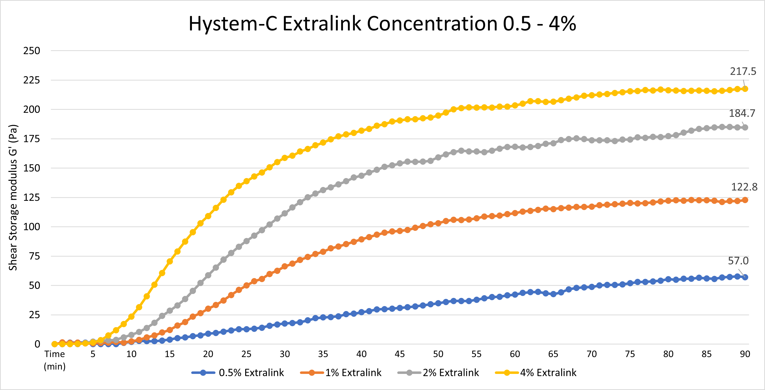
Data was collected using an Elastonsens rheometer from Rheolution: www.rheolution.com
Globular particles less than 75 kDa should be able to freely diffuse through a HyStem hydrogel.
When reconstituted using DG water, the pH of each HyStem component will be approximately 7.4-7.6.
One year from the date of receipt, if stored properly.
Any sterile, deionized, degassed water can be substituted for reconstitution. However, in order to ensure accurate and predictable dissolution and gelation times, our DG Water is highly recommended, as it is degassed, blanketed in argon, and has undergone validation testing with each HyStem component.
Gelin-S provides cellular attachment sites when incorporated in the hydrogel. Gelin-S is thiol-modified, denatured collagen I, derived from either bovine or porcine sources. Gelin-S is included in all HyStem-C and HyStem-HP kits.
Gelin-S has been thiol-modified in the same manner as the hyaluronan in Glycosil (or Heprasil), so that it covalently crosslinks with the Extralink in the HyStem hydrogels.
Yes. Peptides that contain a cysteine residue can be used. The cysteine residue must be present for the peptide to be covalently bonded to the hydrogel substrate.
Yes. ECM proteins, such as laminin, collagen, fibronectin, or vitronectin can be non-covalently incorporated into the hydrogel prior to crosslinking.
HyStem hydrogels and sponges differ in hydration and homogeneity. HyStem sponges are typically polymerized hydrogels that are subsequently freeze-dried. The resulting sponge is a fibrous, mesh network with pores and niches that enable cells to infiltrate and adhere. A true HyStem hydrogel is an encapsulating liquid that polymerizes around suspended cells in culture.
No. The compliance of the hydrogels is set by the amount of Extralink crosslinker added, the concentration of Glycosil (or Heprasil) and Gelin-S used, and the ratio of Glycosil (or Heprasil) to Gelin-S. Once this chemical structure of the hydrogel is fixed, it is not altered by prolonged exposure to cell culture medium.
HyStem sponges can be terminally sterilized by E-beam. HyStem hydrogels have not yet been validated for use with E-beam sterilization methods. HyStem hydrogels are not terminally sterilized by gamma irradiation.
Gelation time is affected by multiple aspects of the gel’s composition.
One way to change the gelation time of a hydrogel is to vary the amount of crosslinker used. Gels with a lower amount of Extralink crosslinker will have a longer gelation time than those with a higher amount of crosslinker. Changing the amount of crosslinker will produce slight changes in gelation time.
Gelation time can be dramatically changed by varying the Glycosil (or Heprasil) and Gelin-S concentrations. Concentrated solutions of Glycosil (or Heprasil) and Gelin-S will create a solution with a much shorter gelation time. This can easily be done by reconstituting the components in a smaller volume of DG Water. Alternatively, diluting these components in larger volumes of DG Water will dramatically increase the total time to form the hydrogel.
HyStem Hydrogels are virtually transparent and should not interfere with microscopy.
HyStem hydrogels may generate mild inflammation as part of the body’s natural healing process in response to injury. HyStem hydrogels do not trigger immune response when used in vivo. (These products are not for human use)
HyStem is degraded in vivo by matrix metalloproteinases (collagenases) and hyaluronidases.
Trypsin, Dipase, collagenase, and hyaluronidase have been used to help detach cells from the surface or from within HyStem hydrogels.
In general, the pore size for HyStem-C and HyStem-HP hydrogels is ~17 nm.
We routinely perform Ellmans test on our Thiolated Hyaluronic Acid. Our product falls between 0.55-0.75 umoles/mg, resulting in a degree of thiolation between 20-30%.
Approximate molecular weight: MW 20000±2000
The approximate molecular weight of the thiolated hyaluronic acid is 300 kDa.
The hyaluronic acid has been thiol-modified on the carboxy groups. These active groups react with acrylate groups on PEGDA, resulting in a hydrogel.
We strongly recommend using the HyStem components the same day that they are solubilized, in order to minimize the chance of autocrosslinking. We cannot guarantee the quality of the product past the first day. If experiments need to be done across two or more days, we recommend leaving the solubilized components at room temperature overnight (capped, in the original vials). We do not recommend re-freezing the components once solubilized.
Product Applications
Read our HyStem eBrochure Here
or
Click on the title of the desired protocol to learn more:
2D Cell Growth on HyStem Hydrogels
HyStem 3D Cell Encapsulation for Cell Delivery Applications Guide
HyStem 3D Cell Encapsulation in hydrogels using 96-well plates
HyStem 3D Cell Encapsulation in hydrogels using TC Inserts
Enzyme Digestion of HyStem Hydrogels for Recovery of Encapsulated Cells
Fluorescent Labeling of HyStem Hydrogels
Cell Recovery from Surface of HyStem Hydrogels
HyStem Gelation Time Variation
Product References
References for HyStem®:
Maloney, E. et al. Immersion Bioprinting of Tumor Organoids in Multi-Well Plates for Increasing Chemotherapy Screening Throughput. Micromachines 11, 208 (2020).
Gaetani, R., et al. (2015) Epicardial application of cardiac progenitor cells in a 3D-printed gelatin/hyaluronic acid patch preserves cardiac function after myocardial infarction. Biomaterials 61: 339-348. PMID: 17335875.
Prestwich, G.D., et al. (2007) 3-D culture in synthetic extracellular matrices: new tissue models for drug toxicology and cancer drug discovery. Adv Enzyme Regul 47: 196-207. PMID: 17335875.
Shu, X.Z., et al. (2006) Synthesis and evaluation of injectable, in situ crosslinkable synthetic extracellular matrices for tissue engineering. J Biomed Mater Res A 79: 901-912. PMID: 16941590.
Shu, X.Z., et al. (2003) Disulfide-crosslinked hyaluronan-gelatin hydrogel films: a covalent mimic of the extracellular matrix for in vitro cell growth. Biomaterials 24: 3825-3834. PMID: 12818555.
S. Cai, et al. (2005) Injectable glycosaminoglycan hydrogels for controlled release of human basic fibroblast growth factor.Biomaterials, 26, 6054-6067.
D. B. Pike, et al. (2006) Heparin-regulated release of growth factors in vitro and angiogenic response in vivo to implanted hyaluronan hydrogels containing VEGF and bFGF. Biomaterials, 27, 5242–5251.
G. D. Prestwich, et al. (2007) 3-D Culture in Synthetic Extracellular Matrices: New Tissue Models for Drug Toxicology and Cancer Drug Discovery. invited, Adv. Enz. Res., in press (2007).
X. Z. Shu, et al, (2006) Synthesis and Evaluation of Injectable, In Situ Crosslinkable Synthetic Extracellular Matrices (sECMs) for Tissue Engineering. J. Biomed Mater. Res. A, 79A(4), 901-912.
Shu, X.Z., et al. (2004) In situ crosslinkable hyaluronan hydrogels for tissue engineering. Biomaterials 25: 1339-1348. PMID: 14643608.
Mehra, T.D., et al. (2006) Molecular stenting with a crosslinked hyaluronan derivative inhibits collagen gel contraction. J Invest Dermatol 126: 2202-2209. PMID: 16741511.
Shu, X.Z., et al. (2004) Attachment and spreading of fibroblasts on an RGD peptide-modified injectable hyaluronan hydrogel. J Biomed Mater Res A 68: 365-375. PMID: 14704979.
Ghosh, K., et al. (2007) Cell adaptation to a physiologically relevant ECM mimic with different viscoelastic properties. Biomaterials 28: 671-679. PMID: 17049594.
Product Certificate of Analysis
Product Disclaimer
This product is for R&D use only and is not intended for human or other uses. Please consult the Material Safety Data Sheet for information regarding hazards and safe handling practices.