-
Collagen
-
Type I - Atelocollagen
- PureCol® Solution, 3 mg/ml (bovine) #5005
- Nutragen® Solution, 6 mg/ml (bovine) #5010
- FibriCol® Solution, 10 mg/ml (bovine) #5133
- PureCol® EZ Gel, Solution, 5 mg/ml (bovine) #5074
- PureCol® Lyophilized, 15 mg (bovine) #5006
- VitroCol® Solution, 3 mg/ml (human) #5007
- VitroCol® Lyophilized, 15 mg (human) #5008
-
Type I - Telocollagen
- TeloCol®-3 Solution, 3 mg/ml (bovine) #5026
- TeloCol®-6 Solution, 6 mg/ml (bovine) #5225
- TeloCol®-10 Solution, 10 mg/ml (bovine) #5226
- RatCol® for 3D gels, Solution, 4 mg/ml (rat) #5153
- RatCol® High Concentration, Solution, 10 mg/ml (rat)
- RatCol® lyophilized, 100 mg (rat)
- RatCol® for Coatings, Solution, 4 mg/ml (rat) #5056
- Type I - Insoluble Collagen
- Type I - Bioinks
- Type II Collagen
- Type III Collagen
- Type IV Collagen
- Collagen Standard
- PureCol® Collagen Coated Plates
- Collagen Scaffolds
- Collagen Hybridizing Peptides
-
Type I - Atelocollagen
- Tunable Stiffness
- CytoSoft® Rigidity Plates
-
Bioprinting
- Support Slurry for FRESH Bioprinting
- Collagen Bioinks for Extrusion Bioprinting
- GelMA Bioinks for Extrusion Bioprinting
- Photoinitiators
- Bioinks and Components for DLP Bioprinting
- Bioink Components
- Methacrylated Collagen
- Methacrylated Gelatin
- Methacrylated Hyaluronic Acid
- Diacrylates
- Methacrylated Polysaccharides
-
3D Hydrogels
- Thermoreversible Hydrogel
- Silk Fibroin
-
Type I Collagen for 3D Hydrogels
- PureCol® Solution, 3 mg/ml (bovine) #5005
- Nutragen® Solution, 6 mg/ml (bovine) #5010
- FibriCol® Solution, 10 mg/ml (bovine) #5133
- PureCol® EZ Gel, Solution, 5 mg/ml (bovine) #5074
- VitroCol® Solution, 3 mg/ml (human) #5007
- TeloCol®-3 Solution, 3 mg/ml (bovine) #5026
- TeloCol®-6 Solution, 6 mg/ml (bovine) #5225
- TeloCol®-10 Solution, 10 mg/ml (bovine) #5226
- RatCol® for 3D gels, Solution, 4 mg/ml (rat) #5153
- HyStem® Thiolated Hyaluronic Acid
- Methacrylated Collagen
- Methacrylated Gelatin
- Methacrylated Hyaluronic Acid
- Diacrylates
- Collagen Sponges
- Methacrylated Polysaccharides
- Extracellular Matrices
- HyStem / Hyaluronic Acid
-
Adhesion Peptides / Proteins
-
Recombinant Adhesion Proteins
- CD2, 0.5 mg/ml #5086
- CDH3, 0.5 mg/ml #5124
- CDH13, 0.5 mg/ml #5125
- CD14, 0.5 mg/ml #5089
- CDH18, 0.5 mg/ml #5090
- CD40, 0.5 mg/ml #5093
- CD86, 0.5 mg/ml #5096
- CD164, 0.5 mg/ml #5100
- CD270, 0.5 mg/ml #5127
- CD274, 0.5 mg/ml #5126
- CD276, 0.5 mg/ml #5123
- E-Cadherin (CD324), 0.5 mg/ml #5085
- ICAM2, 0.5 mg/ml #5107
- Adhesion Peptides
- Collagen Hybridizing Peptides
-
Recombinant Adhesion Proteins
- Reagents
- Assays
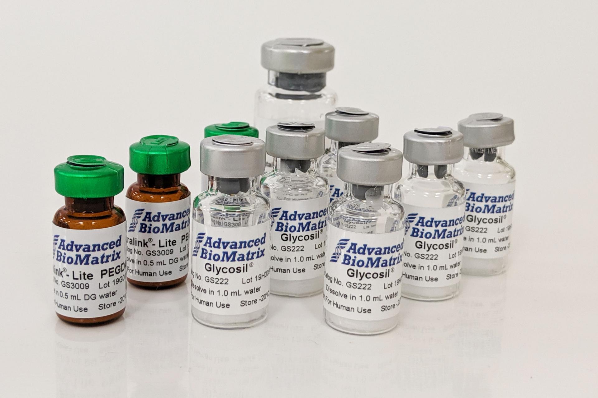
PGSA-INK
5 grams
Catalog #D16110025308
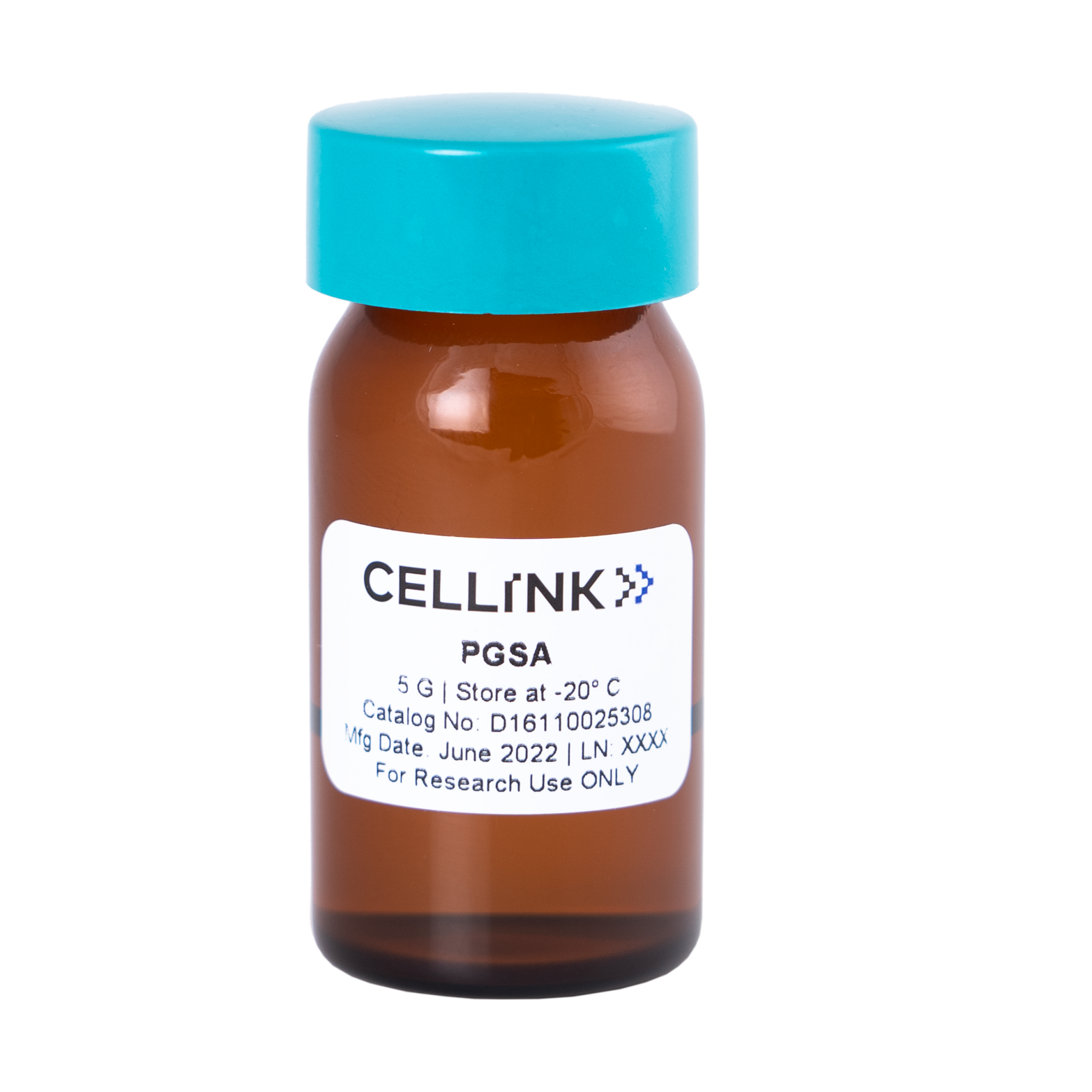
PGSA-INK
5 grams
Catalog #D16110025308
Poly(Glycerol-co-Sebacate) Acrylate - PGSA is 5 grams of photopolymerizable elastomer bioink that can be used for bioprinting and tissue engineering applications with the BIONOVA X. This product has a 6-8 week lead time.
Product Description
PGSA is a bioresorbable polymer with tunable physical properties ranging from an elastomer to a thermoset. It is derived from glycerol and sebacic acid, both of which are naturally occurring metabolites and have been used for biomedical applications regulated by the FDA. In addition, PGSA is light-sensitive and can be crosslinked under exposure of UV and visible light. Due to its great biocompatibility and excellent mechanical properties, PGSA can be used for various applications in tissue engineering and regenerative medicine (e.g. cardiac, nerve, and muscle tissues).
Product References
Salinas CN and Anseth KS, The enhancement of chondrogenic differentiation of human mesenchymal stem cells by enzymatically regulated RGD functionalities. Biomaterials. (2008) 29:2370-7.
Kloxin AM, et al.Photodegradable Hydrogels for Dynamic Tuning of Physical and Chemical Properties Science (2009) 324, 59.
DeForest CA, Polizzotti BD, and Anseth KS, Sequential click reactions for synthesizing and patterning three-dimensional cell microenvironments Nat. Mater. (2009) 8: pp. 659-664.
Baird IS, Yau AY, and Mann BK, Mammalian cell-seeded hydrogel microarrays printed via dip-pin technology BioTech. (2008) 44:249-256.
Baek TJ et al, Photolithographic Fabrication of Poly(Ethylene Glycol) Microstructures for Hydrogel-based Microreactors and Spatially Addressed Microarrays J. Microbiol. Biotechnol. (2007) 17: 1826-1832.
Taite LJ, Rowland ML, Ruffino KA, Smith BR, Lawrence MB, West JL. Bioactive hydrogel substrates: probing leukocyte receptor-ligand interactions in parallel plate flow chamber studies. Ann Biomed Eng. (2006) 34:1705-11.
Du Y, Lo E, Ali S, Khademhosseini A. Directed assembly of cell-laden microgels for fabrication of 3D tissue constructs. Proc Natl Acad Sci U S A. (2008) 105:9522-7.
Khademhosseini A, Yeh J, Jon S, Eng G, Suh KY, Burdick JA, Langer R. Molded polyethylene glycol microstructures for capturing cells within microfluidic channels. Lab Chip. (2004) 4:425-30.
Liao H, Munoz-Pinto D, Qu X, Hou Y, Grunlan MA, Hahn MS. Influence of hydrogel mechanical properties and mesh size on vocal fold fibroblast extracellular matrix production and phenotype. Acta Biomater. (2008) 4:1161-71.
Patel PN, Smith CK, Patrick CW Jr. Rheological and recovery properties of poly(ethylene glycol) diacrylate hydrogels and human adipose tissue.J Biomed Mater Res A. (2005) 73:313-9.
Panda P et al, Stop-Flow Lithography to Generate Cell-Laden Microgel Particles. Lab Chip (2008) 8:1056-1061.
Ma PX and Elisseeff J editors, Scaffolds in Tissue Engineering, CRC Press Boca Raton, FL 2006.
Product Certificate of Analysis
No result for .
Product Disclaimer
This product is for R&D use only and is not intended for human or other uses. Please consult the Material Safety Data Sheet for information regarding hazards and safe handling practices.